Key facts
Most discoid menisci never cause problems and need no treatment. This page is for parents, carers and young people.
- A discoid meniscus is a shock absorber in the knee that is shaped like a disc instead of a crescent.
- It is something a child is born with. It almost always affects the outer side of the knee.
- It is found in about 3 to 5 in 100 people in Western countries, and more often in people of Asian heritage.
- It can cause a clunk or snap in the knee, pain, or a knee that will not straighten.
- If it causes ongoing problems, keyhole surgery can reshape it to a normal shape and repair any tear.
- Surgeons now keep as much of the meniscus as possible. Removing all of it raises the risk of arthritis.
What is a discoid meniscus?
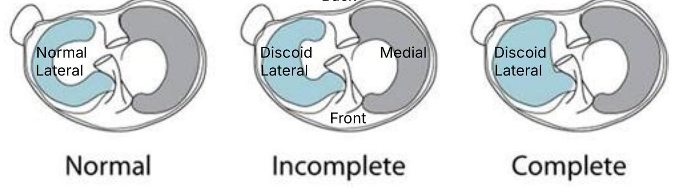
Each knee has two menisci, rubbery pads that sit between the thigh bone and the shin bone. A normal meniscus is C-shaped. A discoid meniscus is wider and often thicker, so it covers more of the shin bone, sometimes all of it.
Doctors describe a discoid meniscus as:
- Incomplete: wider than normal, but not covering the whole outer side of the shin bone
- Complete: covering all, or nearly all, of the outer side of the shin bone
- Unstable: not firmly attached to the edge of the knee, so it can move about. This can happen with either shape.
A discoid meniscus is also built differently inside. Its fibres are less organised and it has a poorer blood supply, so it tears more easily than a normal meniscus.
Who has one and why?
A child is born with a discoid meniscus. It is not caused by anything the child or parents did, and it is not caused by sport.
- It affects about 3 to 5 in 100 people in Western countries, and 10 to 15 in 100 people in East Asia.
- Many people with one never know, because it causes no symptoms.
- It often affects both knees. If one knee causes problems, the doctor will usually check the other knee too.
- Problems usually start in childhood. Younger children tend to have clunking or snapping. Older children and teenagers more often have pain from a tear.
Signs to look for
Symptoms usually start when the meniscus becomes unstable or tears. This often happens without any injury.
Snapping knee (more common in younger children)
- a loud clunk or snap as the knee bends and straightens, which parents may hear or see
- a bulge on the outer side of the knee when it bends
- sometimes no pain at all
Pain or a stuck knee (more common in older children)
- pain on the outer side of the knee
- swelling
- the knee locking or giving way
- not being able to straighten the knee fully, or a limp
- thigh muscles looking thinner on that side
See a doctor if the knee clunks and is painful, swells, locks or will not straighten. A painless clunk on its own usually just needs a check-up.
How is it diagnosed?
- Examination. The doctor checks how the knee moves, listens and feels for a clunk, and looks for a bulge or tenderness on the outer side. Both knees are examined.
- X-ray. The meniscus does not show on X-ray, but there can be clues, such as a wider gap on the outer side of the knee. X-rays also rule out other problems.
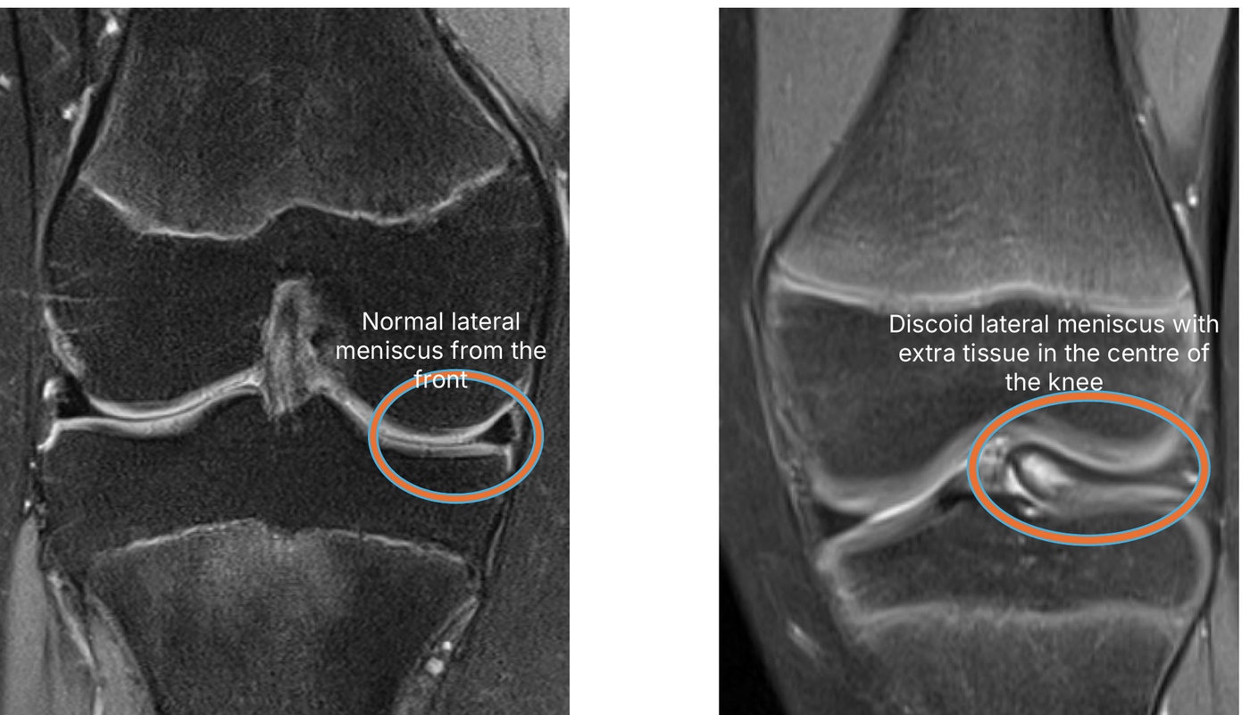
- MRI scan. This shows the shape of the meniscus, any tear, and whether it has moved out of place. It helps the surgeon plan. It does not use radiation.
MRI is not perfect. An incomplete discoid meniscus can look almost normal on a scan. Sometimes the diagnosis is only confirmed during keyhole surgery.
Does it need treatment?
Only if it is causing problems.
| Situation | Usual approach |
|---|---|
| Found by chance, no symptoms | No treatment. Normal activities. See a doctor if symptoms start. |
| Painless clunk only | Usually watched. Doctors are not agreed on whether to operate. |
| Mild or occasional symptoms | Watch, rest and physiotherapy first. Regular check-ups. |
| Ongoing pain, swelling or limits on activity | Keyhole surgery is usually advised |
| Knee locks or will not straighten | Keyhole surgery is usually advised |
There is no benefit in operating on a discoid meniscus that causes no symptoms, including in the other knee.
What does surgery involve?
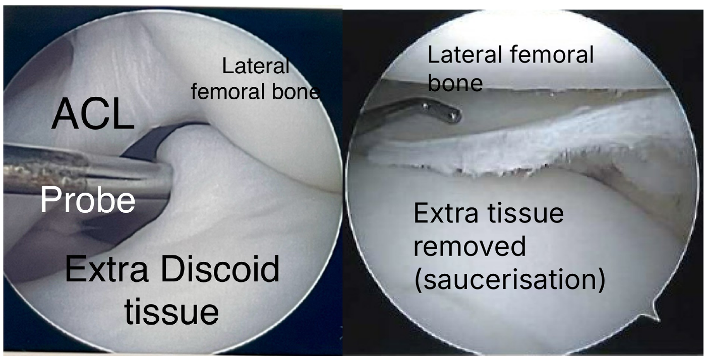
The operation is done by keyhole surgery (arthroscopy) under a general anaesthetic. The aim is to leave a stable meniscus that is as close to normal as possible.
- Look. The surgeon checks the shape of the meniscus, whether it is firmly attached, and whether it is torn.
- Reshape (saucerisation). The extra tissue in the centre is trimmed away, leaving a normal-width rim of about 6 to 8 mm around the edge.
- Repair. Any tear in the remaining rim is stitched.
- Stabilise. If the meniscus is loose, it is stitched back to the edge of the knee.
In the past, the whole meniscus was often removed. This is now avoided, because it leads to more arthritis in the long term. Rarely, if the meniscus is too badly damaged to save, more of it has to be removed.
Recovery and getting back to sport
Recovery depends on what was done. Your surgeon will give you a plan. Typical plans:
| Reshaping only | Reshaping plus repair | |
|---|---|---|
| Walking | Full weight straight away | Crutches with part weight for about 6 weeks |
| Brace | Not usually needed | Hinged brace limiting bending, for about 6 weeks |
| Physiotherapy | From about 2 weeks | From about 2 weeks |
| Return to sport | Gradually from about 8 weeks | Usually from 3 to 4 months, once movement and strength are back |
For children under about 6, a straight knee splint may be used for about 4 weeks after surgery.
Your child should return to sport only when the knee moves fully, is strong, and is not painful or swollen.
Outlook
Most children do well after reshaping, with good knee function and little pain. Repeat clunking is uncommon.
- Results are better in younger children, and when surgery is not delayed for a long time after symptoms start.
- In one study followed for about 10 years, early signs of wear on X-ray were seen in about 1 in 4 knees after reshaping and repair, about 2 in 5 after reshaping alone, and nearly 9 in 10 after most of the meniscus was removed.
- Results can slowly worsen over many years. Long-term check-ups are worthwhile.
- Removing a lot of meniscus can rarely lead to a problem with the bone and cartilage of the outer thigh bone (osteochondritis dissecans).
The other knee may also have a discoid meniscus. It only needs treatment if it starts causing problems.
Making the decision together
Your child should be part of every discussion about their knee. If symptoms are mild, it is reasonable to watch and wait.
Questions you may want to ask:
- Is the meniscus complete or incomplete? Is it stable or loose? Is it torn?
- Are the symptoms bad enough to need surgery, or can we wait?
- Does the other knee have a discoid meniscus too?
- How much of the meniscus will you keep? Will you repair or stabilise it?
- What if you find more damage than expected during surgery?
- How long on crutches, and when can my child play sport?
- How often does your team operate on discoid menisci in children?
About this page
This page gives general information only. It does not replace advice from your child's own medical team.
- Written by: [author name, role]
- Reviewed by: [clinical reviewer, role]
- Next review due: Oct 4, 2027
- Images: used with permission. Patient images are anonymised and shown with consent.
Main sources
- Tapasvi S, Shekhar A, Eriksson K. Discoid lateral meniscus: current concepts. J ISAKOS 2021;6:14-21
- Saavedra M, Sepúlveda M, Tuca MJ, Birrer E. Discoid meniscus: current concepts. EFORT Open Rev 2020;5:371-379
- Kim JH, Ahn JH, Kim JH, Wang JH. Discoid lateral meniscus: importance, diagnosis, and treatment. J Exp Orthop 2020;7:81
- Lee RJ, Nepple JJ, Schmale GA et al. Reliability of a new arthroscopic discoid lateral meniscus classification system. Am J Sports Med 2022;50(5):1245-1253